Home » Hyperspectral Imaging Applications in Biomedical Research
Hyperspectral Imaging Applications in Biomedical Research
Better Imaging
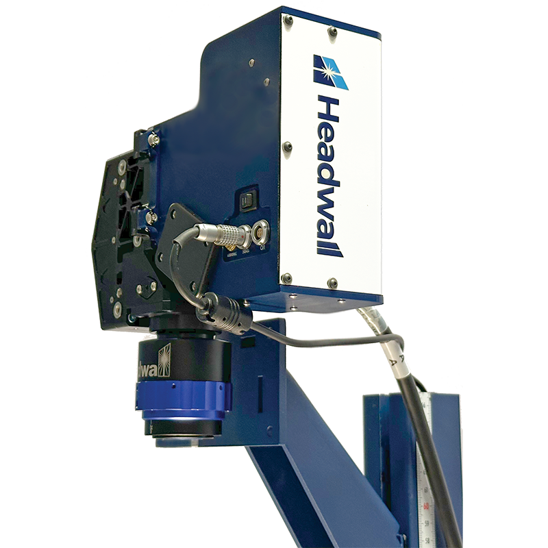
The European collaborative HELICoID project (Hyperspectral Imaging Cancer Detection) uses Headwall’s Hyperspec® imaging sensors to provide real-time identification of tumor margins during brain surgery. It is a non-contact, non-ionizing, and minimally invasive technology that captures image data across hundreds of narrow, contiguous spectral bands.
Screening for a range of neurodegenerative diseases could be a breakthrough benefit of hyperspectral imaging. Patients would undergo a very rapid, non-invasive scan to detect cognitive impairment. With Headwall’s patented technology, the medical community may be able to obtain a highly resolved view of the onset of these diseases at a very early stage.
Improved Surgical Visualization
Real-time identification of tumor margins helps surgeons extract the entire tumor, sparing as much of the healthy tissue as possible. Hyperspectral imaging is a non-contact, non-ionizing and minimally-invasive sensing technique that Headwall has helped pioneer within the European HELICoiD project.
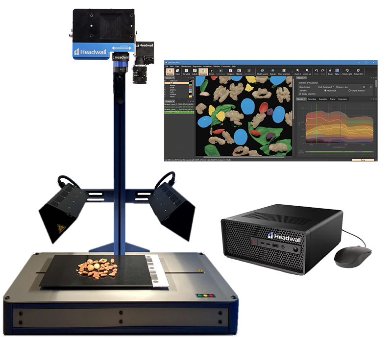
Lab Instrumentation for OEMs
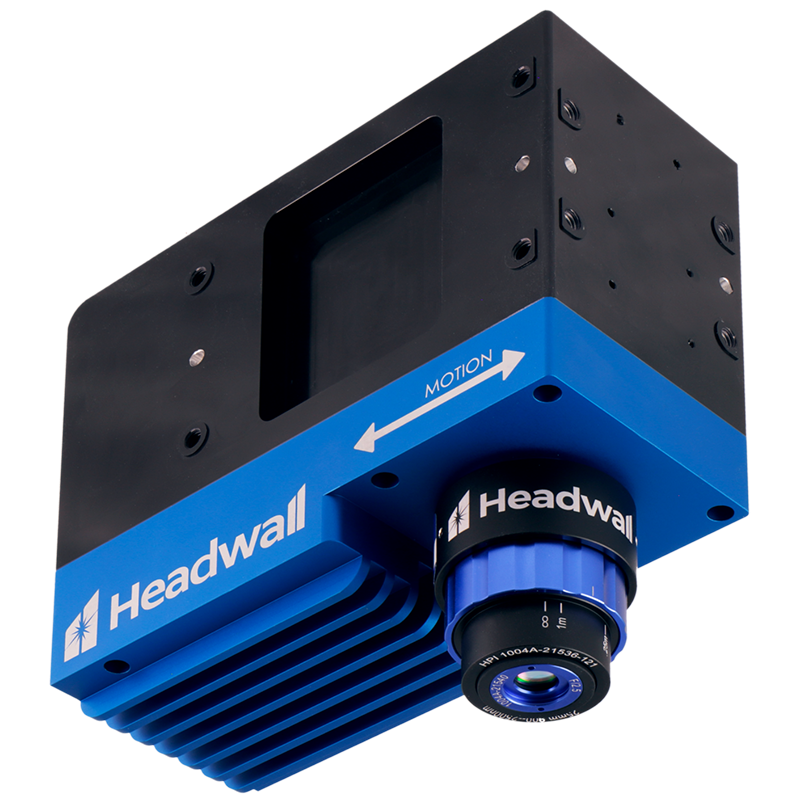
Hyperspectral imaging combines imaging and spectroscopy. With it, every spatial pixel has an entire spectrum. The advantage of collecting unique spectral signatures means that signals having overlapping emission spectra can be distinguished with high accuracy.
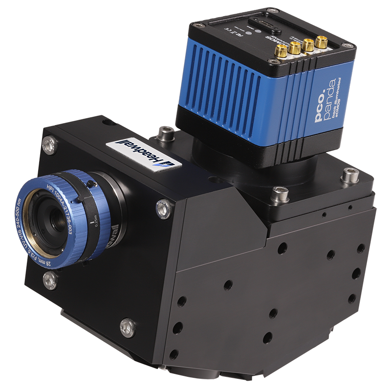
Hyperspectral Microscopy
The emission or reflectance spectrum of a biological sample contains important structural, biochemical or physiological information. Headwall provides the medical-biotech community with advanced imaging solutions that combine high-resolution microscopes and spectrometers. High-throughput screening experiments targeting specific spectral bands are done easily.